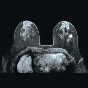
Breast MRI is not a replacement for mammography or ultrasound imaging, but rather is a supplemental tool for detecting and staging breast cancer and other breast abnormalities. MRI is used to:
- evaluate abnormalities detected by mammography.
- identify early breast cancer not detected through other means, especially in women with dense breast tissue and those at high risk for the disease.
- determine the integrity of breast implants.
- distinguish between scar tissue and recurrent tumors.
- assess multiple tumor locations.
- check the progress of chemotherapy.
- look for multiple tumors prior to breast conservation surgery.
- determine whether cancer detected by mammography or ultrasound has spread further in the breast or into the chest wall.
- determine how much cancer has spread beyond the surgical site after a breast biopsy or lumpectomy.
- provide additional information on a diseased breast to make treatment decisions.
Because the risks to a fetus are unknown, pregnant women should not have an MRI exam unless the potential benefit from the MRI is determined to outweigh the potential risks.
For more information on breast MRI visit radiologyinfo.org (Breast MRI).