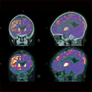
Positron emission tomography, also called PET imaging or a PET scan, is a diagnostic examination that involves the acquisition of physiologic images based on the detection of radiation from the emission of positrons.
Positrons are tiny particles emitted from a radioactive substance administered to the patient. The subsequent images of the human body developed with this technique are used to evaluate a variety of diseases. PET scans are used most often to detect cancer and to examine the effects of cancer therapy by characterizing biochemical changes in the cancer. These scans can be performed on the whole body. PET scans of the heart can be used to determine blood flow to the heart muscle and help evaluate signs of coronary artery disease. PET scans of the heart can also be used to determine if areas of the heart that show decreased function are alive rather than scarred as a result of a prior heart attack, called a myocardial infarction. Combined with a myocardial perfusion study, PET scans allow differentiation of nonfunctioning heart muscle from heart muscle that would benefit from a procedure, such as angioplasty or coronary artery bypass surgery, which would reestablish adequate blood flow and improve heart function. PET scans of the brain are used to evaluate patients who have memory disorders of an undetermined cause, suspected or proven brain tumors or seizure disorders that are not responsive to medical therapy and are therefore candidates for surgery.
For more information on the PET scans visit radiologyinfo.org (PET).